Analysis and Visualisation Software / BESA EEG/MEG Analysis
BESA EEG/MEG Analysis
Versatile and user friendly signal processing for EEG and MEG data. The most widely used software in EEG and MEG research, developed on the basis of 20 years experience in human brain mapping. Wide variety of source analysis algorithms. Bidirectional connection with BrainVoyager™ allows source seeding from fMRI clusters with one mouse click


Click the links below to jump to the relevant section.
Data review and processing module
Source montages and 3D whole-head mapping
ERP analysis and averaging
Source coherence and time-frequency display
Integration with MRI and fMRI
Data Review and Processing Module
This main BESA window provides many tools for reviewing and processing of your EEG or MEG data.
Raw data are directly read using readers implemented for many EEG and MEG file formats. Data processing steps include digital filtering,
artifact detection and correction, computation of correlation and spectral analysis. All these steps can be performed easily with a few mouse clicks.
A variety of different display options allows for convenient review of your data.


Source Montage and 3D Whole-Head Mapping
New montage editor
- Graphical editing of user montages for convenient data review
- Virtual montages with standardized electrode locations
- Combined montages of recorded, virtual, and source channels
- Immediate resorting for regional and hemispheric comparison
Source montages
- Transformation of surface EEG or MEG into brain source activity
- Montages derived from multiple dipole or regional source models
- Standardization for various brain regions or definition by the user
- Additional channels to display PCA components and / or eye artifacts
3D whole-head mapping
- Whole-head spline interpolation for voltage and CSD mapping
- 3D or 2D view of maps, sensors, and head surface points
- Mapping of FFT power, amplitude, and phase
- MEG maps of flux and planar gradients at the scalp surface
- Time series of maps with easy selection of viewpoint, number of maps, and epoch of interest


ERP Analysis and Averaging
This module assists in extracting event-related potentials or fields from your raw data.
It provides an easy-to use interactive tool for designing your own scripted paradigms with predefined trigger definitions, conditions and settings for averaging. Data Files can be scanned automatically for artifacts, and bad channels and samples can be easily identified and excluded using an advanced 2D selection tool. A variety of displays is at hand to view the event-related signals,including 3D whole head maps and event-related (de)synchronization


Source Analysis and Imaging
BESA has a highly interactive and easy-to-use window for source analysis. All relevant information is available at one glance including data, PCA, source waveforms and source localization in 3D head schemes or standardized MRI. You can choose among a variety of state-of-the-art source modeling techniques and head models. 2D and 3D source imaging can be performed with projection onto a standardized MRI.


Source Coherence and Time-Frequency Display
Source coherence analysis reveals the functional connectivity between brain regions. This is achieved by transforming the surface signals into brain activity using brain source montages derived from multiple source models. The new Source Coherence Module provides an extremely fast and user-friendly implementation of time-frequency analysis based on complex demodulation. Users can create event-related time-frequency displays of power, amplitude, or event-related (de)synchronization and coherence for the current montage using brain sources or surface channels. Induced and evoked activities can be separated.


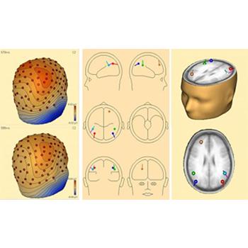
Integration with MRI and fMRI
For an easy superposition of the results of source analysis with individual MRI and fMRI data, BESA provides an interactive link to Rainer Goebel's BrainVoyager program. The bidirectional connection of the two programs allows for source seeding from fMRI clusters with one mouse click.
BrainVoyager™
- Direct and easy interactive user interface of BESA with the BrainVoyager™ program
- Analysis of individual MRI and fMRI data in BrainVoyager™
- Separate license required for the BrainVoyager™ (BV) program.
- MRI and fMRI analysis in BrainVoyager™
- Visualization and processing of individual MRI and fMRI
- Automated rendering of scalp and cortical surfaces
- Expansion and flattening of the cortical surface
- Coregistration of EEG & MEG with MRI BrainVoyager™
- Coregistration of coordinate systems by fiducials and / or surface points
- Direct projection of BESA source models into the individual MRI via interactive link with BrainVoyager™
- Projection of BESA source models into the individual MRI in BESA
- Direct imaging of 3D source images in the individual MRI
- Minimum norm current image based on individual gray/white matter boundary
- Seeding of sources into BESA from anatomical 2D or 3D MR images or from fMRI BOLD clusters in BrainVoyager™ via interactive link
- Overlapped display of fMRI and EEG / MEG sources in BrainVoyager™